Full HTML
Chylous ascites associated with primary pulmonary hypertension: A case report and literature review
Ammar Khan1, Abdo Qaid Lutf2, Ahmed Mahmoud Mostafa3
Author Affiliation
1Medical Student, Department of Medicine, Weill Cornell Medicine, Cornell University,
2Senior Consultant, Department of Medicine, Rheumatology Division, Alkhor Hospital, Alkhor,
3Consultant, Department of Family Medicine, Al Rayyan Health Center, Doha, Qatar
Abstract
Chylous ascites is a rare condition that results from the accumulation of lymph in the abdominal cavity through a variety of mechanisms. We hereby report a case of chylous ascites in a 67-year-old woman who was admitted to our hospital with abdominal distension, bilateral lower extremity edema, and shortness of breath on exertion. The patient had a long history of primary pulmonary hypertension, cor pulmonale, and diabetes mellitus. Imaging studies revealed massive peritoneal fluid, which, after drainage, was found to be consistent with chylous ascites. The patient received high doses of diuretics with a marked improvement in symptoms over the following days. There was no reaccumulation of ascites by the time of discharge. To the best of our knowledge, there are few cases in the literature where chylous ascites has been reported in association with primary pulmonary hypertension.
DOI: 10.32677/yjm.v2i2.3664
Keywords: Chylous ascites, Diuretics, Pulmonary hypertension, Right ventricular failure
Pages: 105-107
View: 6
Download: 13
DOI URL: https://doi.org/10.32677/yjm.v2i2.3664
Publish Date: 28-09-2023
Full Text
INTRODUCTION
Chylous ascites is a milky-appearing peritoneal fluid rich in triglycerides (>200 mg/dL) resulting from the presence of intraperitoneal lymphatic fluid (thoracic or intestinal) [1,2]. It is a rare clinical condition usually caused by malignancy, cirrhosis, infectious causes such as tuberculosis, or postoperative lymphatic disorders in adults [2]. Although rare, cardiovascular diseases such as constrictive pericarditis [3], dilated cardiomyopathy [4], left ventricular heart failure [5], and ischemic heart disease [6] have sometimes been associated with chylous ascites. In this report, we describe a rare case of primary pulmonary hypertension presenting with chylous ascites.
CASE REPORT
A 67-year-old Jordanian female patient was admitted to our hospital with a 24-day history of abdominal distension, bilateral lower limbs edema, and shortness of breath on exertion. The patient had a history of primary pulmonary hypertension, cor pulmonale, and diabetes mellitus for a long time. She denied any surgeries. Her current medications include digoxin 0.125 mg once daily, sildenafil 50 mg once daily, frusemide 40 mg twice daily, and insulin.
On admission, the patient was alert and oriented. Her blood pressure was 140/90 mmHg, her pulse rate was 70 beats/min, and she weighed 88 kilograms. Cardiopulmonary examination showed jugular venous distension, a holosystolic murmur at the left lower sternal border, and basal crackles. The abdomen was markedly distended, with taut skin and a circumference of 82 cm. There was shifting dullness and fluid waves evident on palpation. Bilateral lower limb pitting edema was noted.
Her laboratory tests showed that the hemoglobin level, white blood cell, and platelet count were 11.3 g/dL (normal range, 13 to 16 gldL), 5500/uL (normal range, 4000 to 10000/µL), and 210,000/uL (normal range, 150,000 to 450,000/µL), respectively. The liver function tests disclosed an aspartate aminotransferase level of 31 IU/L (normal range, 0 to 37 IU/L), an alanine aminotransferase level of 38 IU/L (normal range, 0 to 40 IU/L), an alkaline phosphatase level of 254 IU/L (normal range, 40 to 130 IU/L), and a total bilirubin level of 13 µmol/L (normal range, 2.0 to 17.0 µmol/L) respectively. Albumin level was 3.8 g/dL (normal range, 3.2 to 5.2 g/dL), amylase level was 75 IU/L (normal range, 28 to 100 IU/L) and a lipase level was 47 IU/L (normal range, 13 to 60 IU/L). Initial chest x-ray disclosed cardiomegaly but no pleural effusion. Echocardiography showed dilated cardiac chambers, severe mitral, tricuspid, and aortic regurgitation, and the hepatic veins were distended. Pulmonary artery pressure was 75 mmHg and the ejection fraction was 50%. Computed tomography of the abdomen showed massive intraperitoneal fluid, no abnormal masses or enlarged lymph nodes.
Abdominal paracentesis revealed milky fluid (Figure 1) with a high triglyceride level of 210 mg/dl and albumen of 1.7 g/dl, which is consistent with chylous ascites. The white blood cell count was 155/µL. A pigtail catheter was inserted for continuous drainage of ascitic fluid. Investigations to look for the etiology such as chronic liver disease, liver cirrhosis, malignancy, and infection were all negative, and consequently, we concluded that the chylous ascites was caused by right heart failure. Her dietary fat intake was restricted and she received high-dose intravenous diuretic therapy (80 mg twice daily). Over the next few days, her symptoms improved and her ascites volume and triglyceride concentration both decreased. The pigtail catheter was removed, and there was no reaccumulation of ascites by the time of discharge.
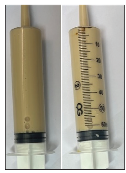
Figure 1: milky ascitic fluid aspirated from the patient
DISCUSSION
The most important step after confirming the diagnosis of chylous ascites is identifying the underlying etiology since prognosis and response to treatment depend on the disease causing it. In our patient, the diagnosis was based on the detection of ascitic fluid with a high triglyceride level (> 200 mg/dL), while pulmonary hypertension was suggested as the underlying etiology after excluding the most common causes of chylous ascites such as disseminated neoplasia and lymphoma, traumatic or surgical rupture of lymphatic vessels and cirrhosis of the liver. Moreover, the occurrence of ascites was noted after diagnosis of heart failure supporting our view in considering pulmonary hypertension as the main culprit. Chylous ascites in the setting of primary pulmonary hypertension most likely occur due to right heart failure. We find no report of chylous ascites due to pure pulmonary hypertension. The only few case reports that we can find in the literature involved right heart failure and pulmonary hypertension simultaneously [7-9].
Chylous ascites has rarely been reported as a consequence of severe right heart failure [10]. The proposed underlying mechanism describes the pathologic events as follows: High venous pressure increases abdominal lymph production secondary to increased capillary filtration. The access lymph is drained through the thoracic duct at up to 12 times the speed. However, lymph flow is limited by the stiffness of the veno-lymphatic junction and the high pressure in the left subclavian vein in the neck. As a result of the restricted lymph drainage, lymph-venous collaterals are formed, which, however, cannot cope with the increased lymph flow. The chylous fluid leaks into the peritoneal cavity or produces protein-losing enteropathy as a result of the rupture of dilated intestinal lacteals [5,10-12].
The standard treatment for chylous ascites has not yet been established. However, the best results have been achieved by treating the underlying cause. In our case, the response to diuretics supports our contention that the underlying etiology of chylous ascites in our patient was right ventricular failure secondary to pulmonary hypertension.
CONCLUSION
In conclusion, we describe a very unusual presentation of pulmonary hypertension. The disappearance of chylous ascites after initiation of diuretic therapy strongly suggests a cause-and-effect relationship between right heart failure and chylous ascites. Physicians should therefore be aware of chylous ascites as a rare manifestation and complication in patients with pulmonary hypertension.
CONSENT FOR PUBLICATION
Written informed consent was obtained from the patient for publication of this case report and all accompanying images.
AUTHORS’ CONTRIBUTIONS
All authors contributed to the completion of this work. The final manuscript was read and approved by all authors
References
- Bhardwaj R, Vaziri H, Gautam A, et al. Chylous Ascites: A Review of Pathogenesis, Diagnosis and Treatment. J Clin Transl Hepatol. 2018 Mar 28;6(1):105-113
- Cárdenas, A. Chylous ascites. The American Journal of Gastroenterology. 2002;97(8), 1896-1900.
- Güneri S, Nazli C, Kinay O, et al. Chylous ascites due to constrictive pericarditis. Int J Card Imaging. 2000 Feb;16(1):49-54.
- Hurley MK, Emiliani VJ, Comer GM, et al. Dilated cardiomyopathy associated with chylous ascites. Am J Gastroenterol. 1989 Dec;84(12):1567-9.
- Ridruejo E, Mandó OG. Chylous ascites as the main manifestation of left ventricular dysfunction: A case report. BMC Gastroenterology. 2005;5(1).
- Levy P, Abadia R, Christoforov B, et al. Chylous ascites complicating ischemic cardiopathy. Presse Med 1985;14:1291. [Abstract]
- Alvarez AB, Ibrahim S, Saggar R. Chylous Ascites in a Case With Right Heart Failure Secondary to Pulmonary Hypertension. Chest. 2012 Oct 1;142(4):130A.
- Zeng W, Hu Y, Feng J, et al. Chylous ascites following repair of total anomalous pulmonary venous connection coexisting with a persistent left superior vena cava in a neonate: a case report. Transl Pediatr. 2021 Jan;10(1):188-193.
- Dash D, Jaladi HC, Nakka S, et al. An Uncommon Cause of Chylous Ascites in an Infant. Indian J Pediatr. 2022;89:1268
- Villena V, De Pablo A, Martin-Escribano P. Chylothorax and chylous ascites due to heart failure. 1995;Eur Respir J 8: 1235-1236
- Dumont AE, Clauss RH, Reed GA, et al. Lymph drainage in patients with congestive heart failure: comparison with findings in hepatic cirrhosis. N Engl J Med 1963; 269: 949–952.
- Wilkinson P, Pinto B, Senior JR. Reversible protein-losing enteropathy with intestinal lymphangiectasia secondary to chronic constrictive pericarditis. N Engl J Med 1965; 173: 1178–1181