Full HTML
Bullous systemic lupus erythematosus
Gerardo Rivera-Silva1, Pablo Martínez-Fernández1
Author Affiliation
1Medical Scientist, Department of Basic Sciences, School of Medicine, University of Monterrey, Monterrey, México
Abstract
A 25-year-old male presented with vaguely painful and pruritic vesicles and bullae on the oral cavity, abdomen, and superior limbs with symmetrical dissemination, which appeared over the past 3 weeks. No clinical history of importance.
DOI: 10.32677/yjm.v2i1.3789
Pages: 58-59
View: 9
Download: 16
DOI URL: https://doi.org/10.32677/yjm.v2i1.3789
Publish Date: 10-05-2023
Full Text
A 25-year-old male presented with vaguely painful and pruritic vesicles and bullae on the oral cavity, abdomen, and superior limbs with symmetrical dissemination, which appeared over the last 3 weeks. No clinical history of importance. The physical examination showed a pallid and prostrated patient, with arterial hypertension (190/110). With several symmetrical dermal vesicles and bullae located on the abdomen and superior limbs and on the oral cavity varying from 1.5 mm to 4 cm (Figure 1).
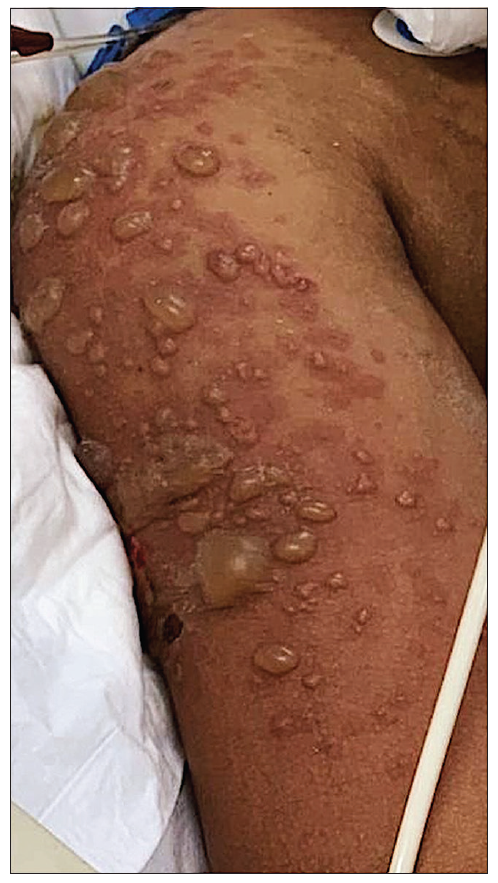
Figure 1: Bullous lesions of varying sizes over erythematosus macules on the right shoulder and upper arm
No lymphadenopathy and sigs of arthritis were identified. Laboratory tests revealed a complete blood cell count with hemoglobin levels of 10.5 g/dL (12.7-15.7 g/DL), leukocytes of 2956 per mm3 (4.3-10.7 x 10³) and platelet count of 120,000 per mm3 (150-350 x 103). Urinalysis shown proteinuria, hematuria, and leukocyturia. The direct Coombs assay, anti-nuclear antibodies, anti-double -stranded DNA and anti-histone were positive. Histopathological analysis reported subepidermal discontinuity associated with neutrophilic infiltrate and direct immunofluorescence was positive for IgG, staining the basement membrane. Elisa was positive for antibodies against type VII collagen. The diagnosis was bullous systemic lupus erythematosus (BSLE) and he was treated successful with rituximab due to poor response to steroid use.
BSLE is an infrequent illness, with an incidence of 3.4 case per million per year, which predominantly develops in females, and less habitually in males [1]. This malady is characterized by vesicles and bullae located on superior trunk, lips, and superior limbs. With symmetrical distribution. While mucosal lesions are usual on pharyngeal, laryngeal, and genital regions that progress with no scratching [2]. Our patient responded satisfactorily to rituximab therapy [3].
Learning points
- Patient education is fundamental in the treatment of BSLE.
- BSLE is an autoimmune disease with various multiorgan involvements, it is crucial to avoid frequent complications include infection, medication side effects and sloughing esophagitis.
- BSLE infrequently recurred.
CONSENT FOR PUBLICATION
Written informed consent was obtained from the patient for publication of this case report and all accompanying images. The patient understands that while every effort is made to maintain the confidentiality of their identity, names, and initials, anonymity cannot be guaranteed.
AUTHORS’ CONTRIBUTIONS
All authors contributed to the completion of this work. The final manuscript was read and approved by all authors
References
- Padrão EMH, Teixeira LF, Maruta CW, et al. Bullous systemic lupus erythematosus a case report. Autops Case Rep. 2019;9(1):e2018069.
- Contestable JJ, Edhegard KD, Meyerle JH. Bullous systemic lupus erythematosus: a review and update to diagnosis and treatment. Am J Clin Dermatol. 2014;15(6):517-24.
- Duan L, Chen L, Zhong S, et al. Treatment of Bullous Systemic Lupus Erythematosus. J Immunol Res. 2015;2015:167064.