Full HTML
An epidural collection due to streptococci Agalactiae
Abdelsalam Abograra, Elmukhtar Habas
Author Affiliation
1Consultant, Department of Radiology, Rhamellia Hospital, Doha, Qatar,
2Senior Consultant, Department of Medicine, Hamad General Hospital, Doha, Qatar
Abstract
An Indonesian lady aged 52 years old presented to the emergency department with a 2-week history of lower backache. One-week later, she developed urine retention followed by bilateral lower limb weakness, and since then, she has been unable to walk. Her medical history, family history, and social history were unremarkable.
DOI: 10.32677/yjm.v1i1.3422
Pages: 52-52
View: 7
Download: 12
DOI URL: https://doi.org/10.32677/yjm.v1i1.3422
Publish Date: 28-03-2025
Full Text
She has no previous history of trauma or similar presented symptoms. Clinical examination showed spastic paraparesis with hyperreflexia. Blood chemistry showed HbA1c of 11.6, and the fasting blood glucose was 14.2 mmol/l. Contrast-enhanced magnetic resonance imaging (MRI) showed an epidural collection extending from T9 to S1 and occupying predominantly the anterior epidural space, with extension toward the posterior epidural space in the lumbosacral region (Fig. 1a). The provisional diagnosis was Pott’s disease, and lumbar (L) hemilaminectomy at L2 was done to drain the epidural collection. Mycobacterium tuberculosis was not detected by acid-fast bacilli or polymerase chain reaction testing of the specimen. However, the drained epdural collection was positive for penicillin-susceptible streptococci Agalactiae. Blood cultures were negative, and transesophageal echocardiography did not show any vegetations. The patient received intravenous ampicillin for 2 weeks, then switched to oral antibiotics for another 6 weeks, and was referred to the rehabilitation center, where she improved and was discharged after 8 weeks with a walker. A repeat MRI (Fig. 1b) showed a complete resolution of the previously described epidural collection.
Streptococcus agalactiae, also known as Group B Streptococcus (GBS), has traditionally been associated with neonates and pregnant women. Invasive diseases due to S. agalactiae are increasing in non-pregnant adults, especially with medical underlying conditions such as diabetes mellitus or neoplasia [1,2]. Although S. agalactiae spondylodiscitis is an unusual manifestation of invasive GBS disease in adults, clinicians should be aware of this clinical entity. It usually occurs in patients with underlying conditions and is usually associated with it a source of infection with a favorable outcome [2,3]. Our patient was found to be diabetic and responded adequately to therapy; nevertheless, we could not find the source of the infection.
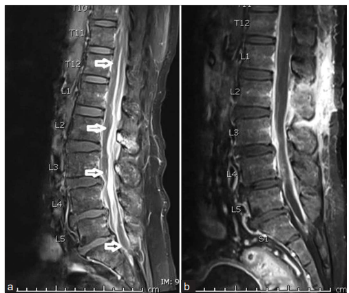
Figure 1. Post-contrast fat-saturated sagital t1-weighted magnetic resonance images (MRI) (a and b) of the spine. Figure 1a shows an epidural collection extending from T9 to S1 and occupying predominantly the anterior epidural space, with extension towards the posterior epidural space in the lumbosacral region (white arrows).(b) Post-therapy MRI shows complete resolution of the previously noted epidural collection with sustained epidural enhancement
References
1. Khan FY. Streptococcus agalactiae meningitis in adult patient: A case report and literature review. Case Rep Infect Dis 2016;2016:6183602.
2. Raabe VN, Shane AL. Group B Streptococcus (Streptococcus agalactiae). Microbiol Spectr 2019;7:10.
3. Martinaud C, Gaillard T, Graffin B, et al. Vertebral osteomyelitis due toStreptococcus agalactiae ST-17. Ann Biol Clin (Paris) 2008;66:87-9.